
This topic is very poorly understood. How does fluence and pulsewidth relate to cellular denaturation – or, ‘cooking’ of the cells.
I’m going to portray this in a series of graphs – I hope this works…

When we fire light energy into the skin, we can generate some heating, if there is something in there to absorb that light energy. In most skin treatments, that is precisely what we are trying to do.
As the fluence at the skin surface is increased, the temperature of any absorbing target in the skin will also increase.
Very simple.
What about the absorption coefficient of the absorbing target (I hear you ask)?

The absorption coefficient of any tissue or cell determines how much light it will absorb. Cells with low coefficients will absorb quite poorly, resulting in low temperature rises. Conversely, any target with high absorption coefficients will rise in temperature much more, for the same incident fluence.
This is also simple!
What happens with different pulsewidths?

Now this does depend on the particular technology, but generally, with continuous lasers, longer pulsewidths will result in higher temperature increases in the skin.
However, for pulsed systems (lasers and IPLs), it is usually the other way round!! Shorter pulsewidths will generate higher temperatures, simply because there is less time for the heat to escape from the absorbing target during the pulse.
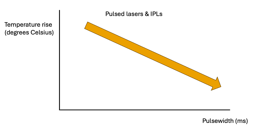
This is a bit more confusing!
OK, so we can see how changing the fluence, absorption coefficient and pulsewidth can change the rise in temperature. Note that these rises are all “linear”. In other words, the longer you apply the energy/fluence, the higher the temperature will rise in a corresponding, straightforward manner.
BUT….
…cell or tissue denaturation (‘cooking’) does not behave like this.

The rate of cell death, due to heating, is exponential – not linear! So, a small increase in temperature can result in a rapid increase in cell death.
This means that increasing the fluence even a relatively small amount, can lead to a significant increase in the rate of cooking.
Plus, this rate also depends on the actual temperature of the cells. So, cells at 85°C will break down much faster than cells at 75°C, and many times faster than cells at 65°C – this is the nature of exponential growth! As a result, the cells ‘cook’ much faster at these higher temperatures.
For example, when considering bulk dermis, at a temperature of 75°C, it will take those cells around 6.75 milliseconds to fully denature. But at 85°C, it only takes 0.29 ms. So, a 10°C rise in temperature, reduces the cooking time by a factor of just over 23.
| Temperature range (°C) | Time to denature (ms) |
| 60 – 65 | 1097 – 191 |
| 80 – 85 | 1.36 – 0.29 |
In the table above we can see that the time required to denature cells in the range from 60 to 65°C is from 1097ms down to 191ms. But, for the range 80 to 85°C, the time required drops from 1.36ms to 0.29ms – significantly shorter! Clearly, the higher the temperature, the faster cells denature.
Now, this is very important because it means that applying a higher fluence to targets like hair or blood vessels, can induce the desired reaction in much shorter timescales. And this is why the pulsewidth becomes less and less important as the fluence increases. With higher cell temperatures, the rate of cooking is so fast, that the pulsewidth becomes irrelevant – particularly at higher temperatures (higher fluences).
This is why I always advocate higher fluences (with corresponding skin cooling) for the treatment of hair and vessels. Then we don’t need to worry about the pulsewidth at all.
Fluence ‘trounces’ pulsewidth – every time!
Hope this helps,
Mike.

P.S. You can now find our podcast at https://podcasts.apple.com/gb/podcast/lasers-in-skin/id1790635555?i=1000691630851, or wherever you usually find your podcasts.


Mike – thanks for the great resources. I came across your first ed book last year and it has been an invaluable resource. Also just started following the PODcasts. I enjoyed the explain it like I’m 5… that’s my level.
I’m an Australian based Dr. We use red / blue light LED PDT extensively for early superficial skin cancers and precancerous lesions.
My question is clinics are purchasing lower cost lamps that don’t quote the fluence. Proxy measurements like lux are being used as a measure of the fluence.
I understand at a simple level what fluence is – the bus analogy – but my question is how is this / can this be measured ?
Many thanks
Hi Chris,
Thanks for your kind comments.
Do you know the fluence of your LED units? Can you let me know which ones you are using and I’ll Google them?
You can measure fluence in one of two ways…
Either, by using an energy meter and taking a bunch of readings and spot diameters. That will be tricky for an LED output though! Alternatively, a meter designed to measure fluence specifically will give you the answer. But these are quite expensive since they are very ‘bespoke’!
I don’t know enough about PDT and the threshold fluences required. I suspect they are quite low, hence the use of LEDs. But they will still need to exceed some threshold!
I hope this helps,
Mike.