
I often hear laser/IPL operators saying that they “don’t want to burn their patients/clients.” They are wrong!! They DO want to ‘burn’ them, but in a very controlled manner…
Every photothermal treatment (the removal of hair, blood vessels, benign pigmentation etc) requires delivering light energy to generate a thermal (heat) response in the tissues. We are literally trying to ‘burn’ the unwanted cells to kill them.
If we apply the correct energy (fluence) in the correct pulsewidth (cooking time) then we can safely and effectively destroy the unwanted cells, without damaging the surrounding tissues.
My calculations reveal that most of the light energy we fire at the skin does NOT do the job we want it to!! In fact, in some circumstances, less than 1% of the light energy is actually doing the job!! That means that more than 99% of the light energy is generating excess heat in the skin, looking to cook something. This is not good…
To mitigate against this we must apply plenty of cooling to the skin. After all, we are trying to ‘burn’ it, but not all of it!
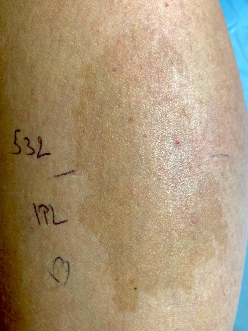
I carried out a wee test to show the difference between treating a benign pigmented lesions with the Q-switched Nd:YAG laser using the 532nm wavelength and an IPL using the 480nm filter.
So, while the laser will only generate a 532nm beam (which is strongly absorbed in both melanin and blood), the IPL will produce a range of wavelengths from 480 up to 1200 nm. Also, the QS laser is fired in an extremely short pulse – only about 10 nanoseconds long, while the IPL was fired in a 35 millisecond pulse (which is 3.5 million times longer!!)
As a consequence of these pulsewidths, the Nd:YAG laser generates a ‘photomechanical’ response in the melanosomes while the IPL produces a ‘photothermal’ reaction. In addition, the strong absorption by the blood coupled with the very short pulses will cause the capillary vessels to burst apart too.
The following set of photos come from a young lady who had a benign pigmented mark on her calf (many thanks to her for her kind permission to use these photos). She agreed to try both the QS 532nm beam and the IPL beam to see how they differed.
We decided to treat the top half with the QS Nd:YAG laser, and the bottom half with the IPL.
The first treatment was done on 26.10.21. I applied a fluence of 2 J/cm2 in a 532nm beam. This photo shows the immediate reaction following treatment by the YAG laser. The melanosomes have absorbed the laser energy which has generated steam bubbles on their surfaces. The whitening reaction is the result of these steam bubbles. This photo was taken before we applied the IPL beam to the lower half.
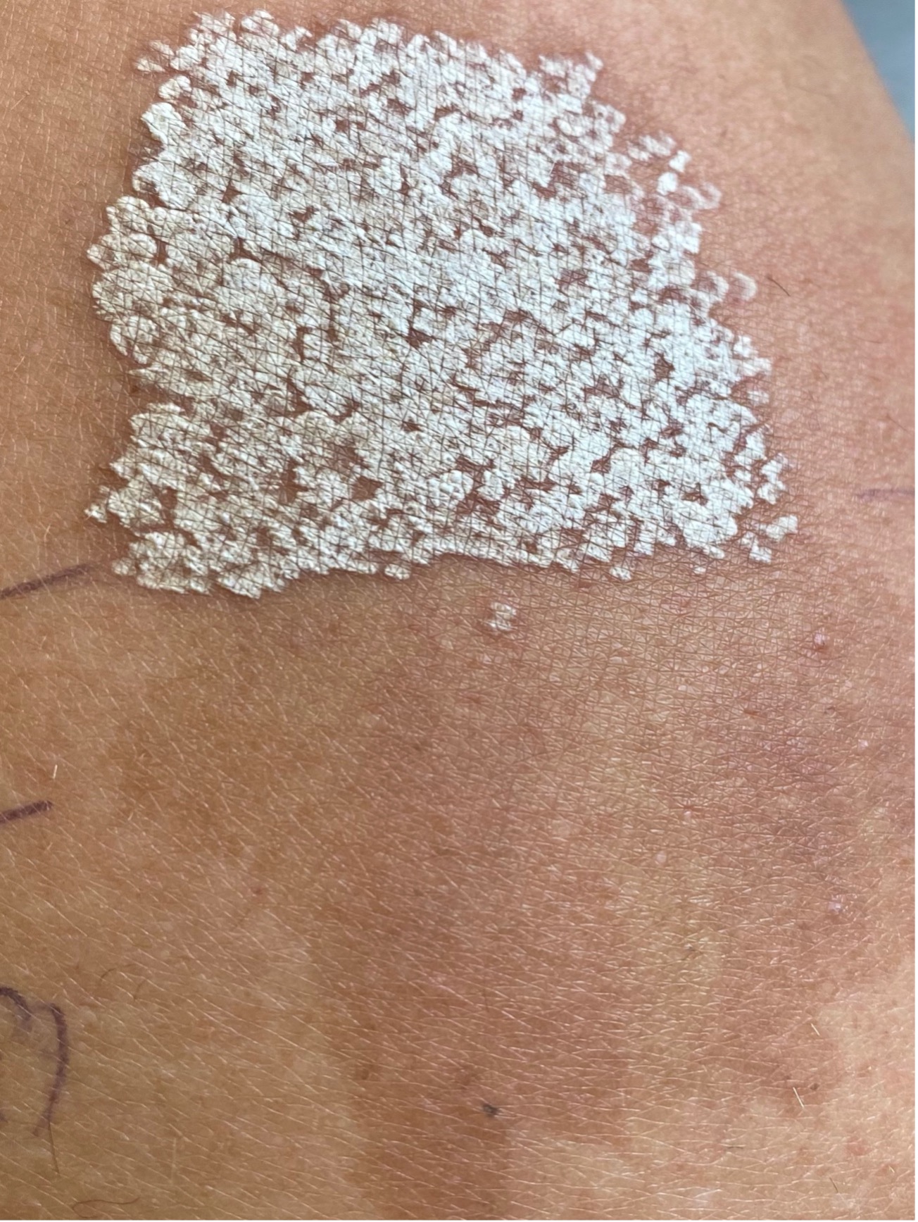
I didn’t take a photo immediately after the IPL treatment – there wasn’t much to see! However, I treated the lower half with a fluence of 20 J/cm2 using a 480 nm filter in a 35ms pulse.
This next photo was taken the next day – 27.10.21:
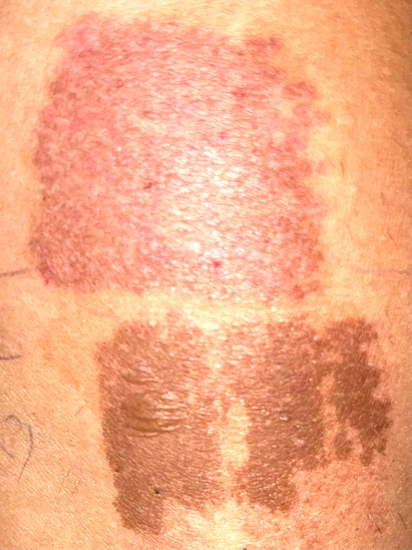
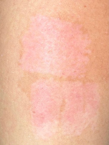
Here we can clearly see two different reactions to the treatments. The upper area has turned a red colour – this is primarily due to the damage to the capillaries induced by the strong absorption of the 532nm wavelength.
The lower area is showing some light blistering where the IPL’s thermal energy has disturbed the melanosomes. The skin has a brown appearance due to melanin’s reaction. Already it appears that the IPL area should have received more cooling immediately after treatment!!
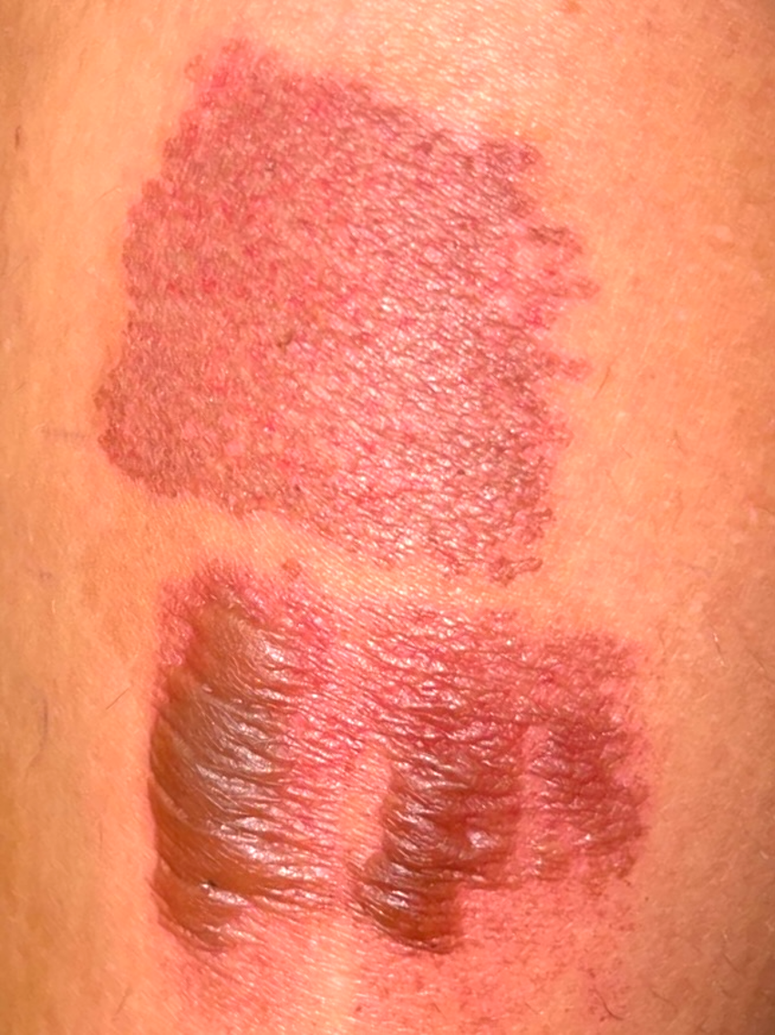
On the following day (28.10.21) the treated areas looked like this:
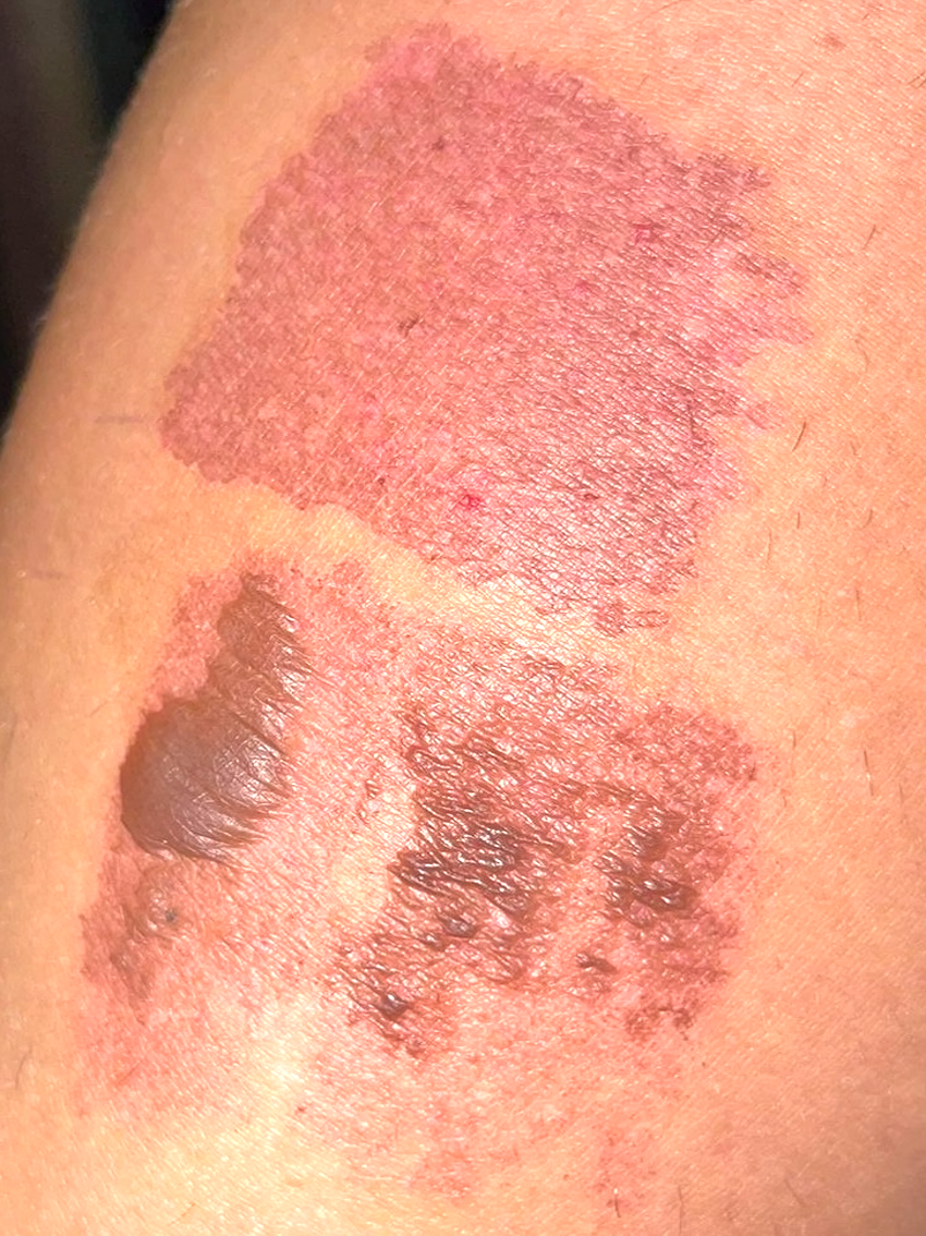
The main blister has increased in size in the IPL area, with a few smaller blisters beginning to make an appearance. The YAG area hasn’t changed much.
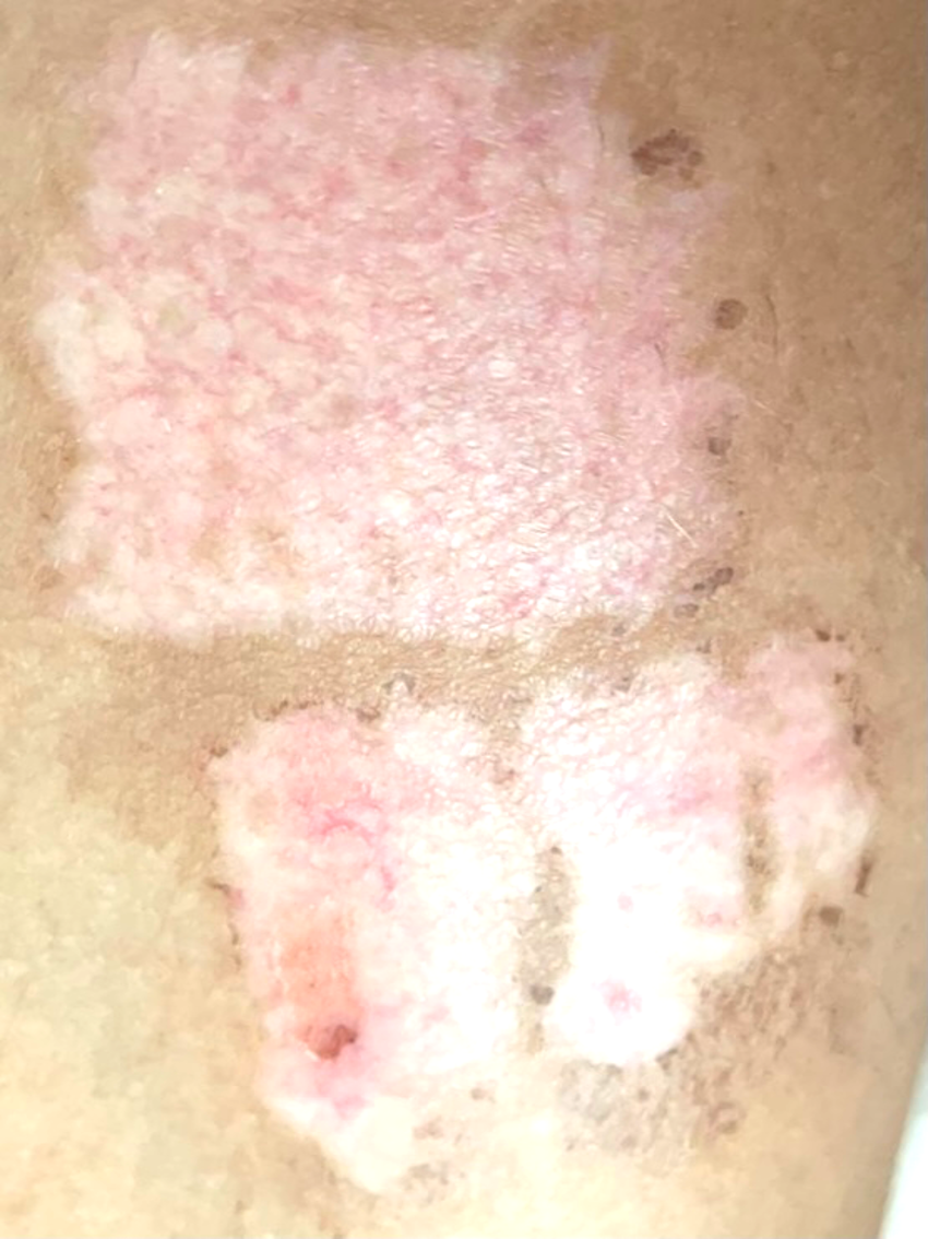
On the following day (29.10.21) the blisters in the IPL area have increased in size. Now the skin looks like it has been ‘burnt’. The patient said that the blisters were “a bit sore and makes sitting a little uncomfortable.”
On the following day (30.10.21) the YAG area has begun to change colour to a brownish tint, while the IPL area is definitely looking damaged!

On the 31.10.21 the blisters have burst naturally and that area began to itch, according to the patient.
On 1.11.21 we can see that both areas appear quite similar and that the blistered tissue is beginning to fall off the skin, exposing the underlying dermis.
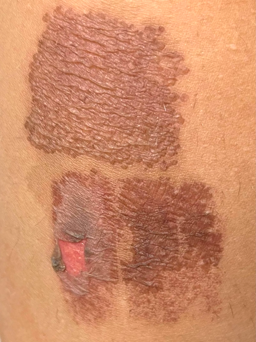
On 2.11.21 (below) we can now see that the blistered tissues are falling away from both treated areas.
There is clearly a little more vascular damage in the IPL area, compared with the YAG area at this stage.
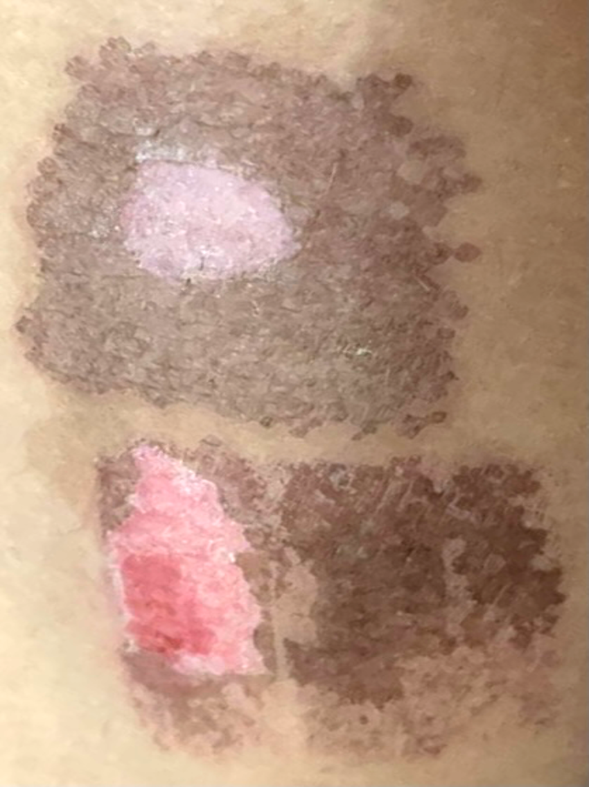
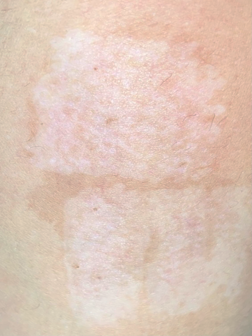
Above is 3.11.21 – 7 days post-treatment. Most of the dead surface tissue has fallen away from the skin in the YAG treated area, while it is beginning to fall away from the IPL treated area below. The underlying dermal tissue appears to be in good condition with no significant signs of damage.
Remember, the blistered tissue contains the melanin, which we were trying to remove. By falling off like this, we have achieved our goal.
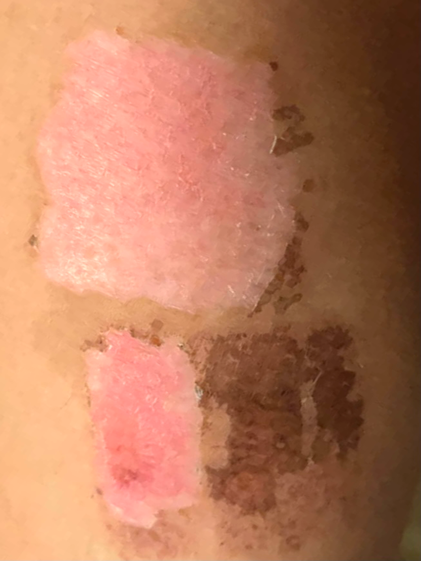
A couple of days later (5.11.21) and we can see the repair processes have definitely begun.
All the blisters have fallen away exposing the underlying dermis, by this point (9 days post-treatment).

A few days later (11.11.21) and we can clearly see the areas that we ‘missed’ in the treatments. The dermis is now looking very healthy and most of the blisters have finally gone.
On 19.11.21 the skin is looking much better with a more ‘normal’ appearance beginning to show.
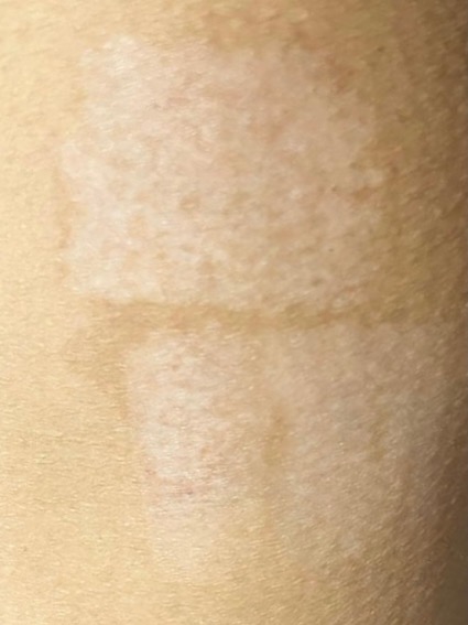
A month after the treatment (27.11.21) and the skin is looking good. The patient had applied fake tan at this point making the appearance much more ‘normal’.
On 9.12.21 both areas now look very similar (44 days after the treatment). It is difficult to tell which area received the YAG laser and which had the IPL treatment! Even though the treatment pathways were quite different, the final results are remarkably similar. This shows that it doesn’t really matter how you kill the unwanted cells, as long as you don’t damage the adjacent tissues (collagen, nerves etc).
Conclusions
As I said at the start, we are deliberately trying to ‘burn’ the skin with photothermal treatments. But these are “controlled” burns, using the correct parameters and protocols. If carried out correctly, we can achieve very nice results with just the right amount of damage, but without scarring.
Always start with low fluences – with both Q-switched (or pico) lasers and IPLs. With such superficial pigment, we don’t need high fluences – they will only end up damaging the dermis with excess heat.
We must also use the appropriate skin cooling techniques to minimise unwanted damage. I use a protocol where I pre-cool the skin before firing the light energy, then ‘post-cool’ immediately, then ‘post-cool’ again using ice-packs. I will soon make a video describing this technique. It helps to both reduce the pain sensation that patients feels and protect the skin form too much thermal damage.
However, we must inform the patients/clients in advance – let them know what to expect or they will likely panic. Feel free to use these photos if you wish to explain what they might expect to see. If they know in advance, they will be much less likely to become worried or upset. Also, make sure they understand how to look after their skin following these treatments.
Hope this helps,
Mike.
You can learn more about the fundamentals of laser/IPLs and their interactions in skin with our free, online eBook. Click here to find out more.
