Are we optimising the wrong part of the tattoo removal process?
In this post I look at picosecond versus nanosecond (Q-switched lasers) evidence, collateral tissue trauma and the biology of pigment clearance, and question if we are approaching this issue properly.
The question behind the technology
For over four decades, developments in tattoo removal have emphasised laser technology: shorter pulses, higher peak irradiance (power density) and greater photomechanical disruption of tattoo pigment. Picosecond lasers are often promoted as “faster” and more “effective” than conventional nanosecond Q-switched systems. But an essential fact risks being overlooked: the laser initiates the overall process; but it is the body that is responsible for much of the subsequent clearance of the ink particles.
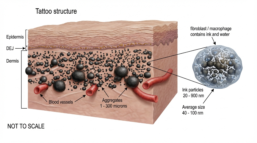
Around three months after ink is injected into the skin, most of the ink particles have formed into aggregates which are either removed to the lymph nodes by lymphatic macrophages, or remain in the dermis within dermal macrophages and fibroblasts.
Laser irradiation can alter pigment particles and pigment-containing cells within an extraordinarily short interval. However, the resulting biological response unfolds over weeks or months. Optimising the first event does not necessarily optimise the whole ink clearance process.
What does the comparative evidence show?
Randomised split-tattoo studies demonstrate that some picosecond systems outperform some nanosecond systems. Other studies, particularly at 1064 nm for black tattoos, have not found a significant difference in clearance. The central issue is what those comparisons can legitimately establish.
755 nm alexandrite: Ma et al. (2026)
A randomised split-tattoo study involving 30 patients reported substantially better clearance with a picosecond alexandrite system after five sessions: median clearance score 8 versus 5, and satisfactory clearance in 73.3% versus 23.3% of treated areas. Complete clearance was reported in only 10% of picosecond-treated areas.
The protocols reportedly used different fluences and sometimes different spot diameters. Published secondary summaries give approximately 2.68–5.25 J/cm² for picosecond treatment and 5–8 J/cm² for nanosecond treatment; these ranges require verification against the original methods before quotation as definitive. The clinical result is noteworthy, but pulse duration was not isolated as the only changing variable. This is critically important! If more than one variable is allowed to change – fluence, spot size and pulse duration – then we cannot assign any significant difference to only one of those variables!! It is simply not possible to know which variable had which effect unless all the others are ‘locked down’. This is basic experimental science.
1064 nm Nd:YAG: Pinto et al. (2017)
This split-tattoo comparison used approximately 450 ps and 5 ns pulses for black tattoos. After two sessions, the investigators found no statistically significant difference in clearance, although reported pain was lower with the picosecond system. Fluences were clinically selected using immediate treatment responses rather than explicitly matched across tattoo halves. Thus, the study compares clinical protocols, not pulse duration alone. As with the Ma study above, it is not possible to pin down any serious conclusion about the effects of pulse duration with these results.
532/1064 nm comparison in Asian patients (2020)
A further prospective comparison reported favourable picosecond outcomes, but at 1064 nm the reported settings were 4.3 J/cm² (450 ps) and 5.0 J/cm² (50 ns), each with a 3 mm spot. Again, fluence and pulse duration differed together.
| Study | Main observation | Interpretive limitation |
| Ma et al., 2026; 755 nm | Greater clearance with picosecond protocol | Fluence and spot size not consistently matched |
| Pinto et al., 2017; 1064 nm | No significant clearance difference after two sessions | Fluences chosen by clinical endpoint |
| Prospective Asian study, 2020 | Picosecond advantage reported | Different fluences at matched 3 mm spot |
These are useful pragmatic comparisons of commercial treatments. They do not, by themselves, demonstrate that pulse duration is the independent cause of superior outcomes. Equally, the use of different fluences does not invalidate the trials: clinicians also need to know which complete protocol works better in practice.
An unexpected natural experiment: the COVID-19 treatment interruptions
In 2024, I published “Longer Intervals Between Sessions With Q-Switched and Picosecond Lasers Result in Enhanced Tattoo Ink Clearance: Case Study Series of 12 Patients” in the Journal of Dermatology Research. The report brought together photographs and treatment histories from 12 patients treated at nine centres in the UK, USA and Canada. Pandemic-related interruptions, alongside other extended gaps, offered an unusual opportunity to observe tattoos long after their last laser exposure.
Several cases were striking. In Case 7, a tattoo showed substantial apparent fading over a 23-month interval following one 1064 nm Q-switched Nd:YAG session (6 ns). Case 9 was photographed 21 months after one 1064 nm treatment (6 ns). Case 12 showed marked change over a 51-month interval after a single 1064 nm session (6 ns). The series also included a 755 nm picosecond alexandrite case, demonstrating that prolonged follow-up is relevant across laser technologies.
These observations challenge the assumption that the biologically meaningful outcome of a session can be assessed fully after only six to eight weeks. They do not, however, show precisely when fading occurred during the long intervals: serial standardised measurements were not available. Nor does a selected case series establish that longer gaps cause greater ultimate clearance, or that every patient benefits from postponing repeat treatment sessions.
From treatment interval to tissue environment
My 2024 paper also considered macrophage-mediated pigment capture and recapture, lymphatic transport and microscopic tissue changes associated with tattooing and repeated laser exposures. It raised the possibility that repeated treatments can add injury to the dermis while an earlier session’s biological response is still evolving. This is the bridge to the present hypothesis: could reducing collateral injury preserve a tissue environment more favourable to continued pigment elimination?
The distinction is essential. The case series supports prolonged apparent clearance after treatment; it did not measure tissue trauma against clearance rate, quantify macrophage trafficking or test low-fluence exposures. Those remain unanswered mechanistic questions. The new hypothesis therefore extends the 2024 observations rather than being proven by them.
It wasn’t until recently (late 2026) that I noticed something else about the result in my 2024 paper – most of those clinical results were observed after a very small number of treatments – typically one or two! I didn’t see similar results in people who had had many sessions…
Two complementary variables to investigate
Treatment timing asks how long the body should be allowed to respond before another exposure. Treatment fluence asks whether the initial irradiation produces enough useful pigment alteration without imposing avoidable tissue injury. A rigorous prospective study could assess both variables independently, then examine whether they interact. Its outcomes should include calibrated pigmentation measurements, adverse tissue responses and long-term follow-up—not just immediate whitening.
A neglected possibility: less trauma, better clearance
Picosecond systems frequently operate at lower incident fluences, in part because of their design and engineering constraints. Could some observed clinical advantages arise not solely from higher peak irradiance, but from a more favourable post-treatment tissue environment – i.e. less trauma?
A higher-fluence treatment may produce substantial pigment alteration, yet also cause oedema, epidermal injury, vascular disturbance, inflammation or, after repeated treatments, fibrosis (scar tissue formation). These responses may alter local transport and tissue organisation during the period in which pigment is being processed. A lower-trauma treatment might preserve a more favourable environment for macrophage activity and subsequent pigment movement.
This is my current hypothesis, not an established mechanism. Inflammation and macrophage recruitment are also part of normal pigment clearance; tissue repair and pigment processing are not necessarily competing for a fixed pool of “resources”. Furthermore, lower incident fluence does not guarantee lower injury when pulse duration, beam profile and absorption differ. Ideally, injury must be measured, not inferred from the fluence setting.
Nevertheless, the possibility is important: additional pigment fragmentation may offer diminishing returns if additional collateral injury compromises the longer-term biological response.
This is the main point of this post – I think we need to consider not only the technology we use, but also the physiological processes post-treatment – over which we have very little control, after the treatment. I now suspect that the latter issue is significantly more important than we had previous thought.
Peak irradiance is not the clinical endpoint
At equal fluence, a 500 ps pulse has ten times the average irradiance during the pulse of a 5 ns pulse. This may change stress generation and pigment disruption within the dermal macrophages. It does not follow that the body will eliminate ten times as much pigment, or eliminate it ten times faster. Why should it?
Clinical lightening itself is an imperfect surrogate for true pigment elimination. Changes in the optical properties of the ink, redistribution and transport to lymph nodes can all influence appearance. A strong immediate whitening response (frosting) is likewise not a quantitative measure of eventual biological clearance. As I have explained previously, frosting may occur in both the ink aggregates and in the epidermal melanin. But I now suspect that frosting might indicate “too much” fluence!!
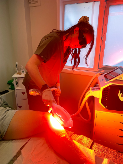
Could there be an optimum fluence?
A useful testable proposition is that clearance versus fluence may be non-monotonic. At very low fluence, pigment alteration may be insufficient. At intermediate fluence, useful alteration may occur with limited dermal injury. At higher fluence, additional tissue trauma might eventually offset the benefits of further pigment disruption. This shape of the response curve has not been established clinically; it is precisely what a properly designed experiment should test.
The important question becomes: what is the minimum physical intervention that reliably initiates effective biological clearance, without unnecessary collateral tissue injury?
How should this be tested?
A controlled study using one nanosecond Q-switched Nd:YAG platform could hold wavelength, pulse duration, spot size and beam profile constant while investigating several fluences. Within-patient comparisons, objective longitudinal photography or spectrophotometry, blinded assessment and extended follow-up would help distinguish early optical change from sustained clearance.
The study should record not just pigment lightening but delayed erythema, oedema, blistering, crusting, healing time and, where feasible, measures of dermal change. The key analysis would compare both the magnitude and kinetics of clearance against the measured burden of tissue injury. Conventional treatment should remain an appropriate comparator.
Very low-fluence exposures, including proposed ranges around 0.1–0.2 J/cm² at 1064 nm, remain experimental: neither sufficient pigment alteration nor clinical efficacy has been demonstrated at those settings. Any human investigation would require suitable ethical review, consent, safety controls and follow-up.
What would count as faster removal?
Greater lightening after a fixed number of sessions is not the same as fewer sessions to near-complete clearance. Nor is it the same as less elapsed time from the first exposure to a satisfactory endpoint. Future trials should report all three, alongside adverse effects and continued lightening after the last session. My 2024 paper shows that ink clearance can continue for up to two years after the last treatment session!
Conclusion: optimise the biology, not just the pulse
Picosecond lasers can offer genuine clinical advantages under particular conditions. But the clinical evidence does not support the simple proposition that shorter pulses must always produce faster or more complete tattoo removal. Where fluence and other variables differ, the contribution of pulse duration alone remains uncertain.
An equally important question has received far less attention: does minimising collateral tissue trauma allow the body to clear altered pigment more effectively? The answer is not yet known. It is biologically plausible, measurable and worth investigating. My 2024 case series offers observational support for the importance of prolonged biological clearance, while the proposed low-trauma mechanism still requires direct experimental testing.
The objective should not necessarily be maximum immediate pigment disruption. It should be maximum safe, sustained biological clearance. And, finally, we must remember that…
“Lasers don’t remove tattoos —
the body does.”
I hope this all makes sense,
Mike.

We will be discussing this further in one of our upcoming podcast episodes…

Selected references and verification notes
- Ma et al. (2026). Randomised split-tattoo comparison of 755 nm picosecond and nanosecond alexandrite lasers. Plastic and Reconstructive Surgery. PMID: 41729109. Verify detailed laser settings in full text before publication.
- Pinto et al. (2017). Comparison of picosecond and nanosecond 1064 nm Nd:YAG lasers for tattoo removal. British Journal of Dermatology. PMID: 27518129.
- Prospective comparison of 532/1064 nm picosecond and nanosecond lasers for professional tattoos in Asian patients (2020). PMCID: PMC7447827.
- Systematic review of picosecond laser tattoo removal (2020). PMID: 32763326.
- Comparative meta-analysis of picosecond versus nanosecond lasers for pigmentation (2025). PMID: 41184656.
- Murphy M. (2024). Longer Intervals Between Sessions With Q-Switched and Picosecond Lasers Result in Enhanced Tattoo Ink Clearance: Case Study Series of 12 Patients. Journal of Dermatology Research. 5(2):1–14. DOI: 10.46889/JDR.2024.5211.